Monday, December 10, 2007

The Case of the Upper Airway Obstruction

Hello once again from the land of veterinary medicine. When I last left off, I detailed my entrance into the infamous block of bad news and promised to tell of my most challenging case yet. Towards the end of the first week in the rotation, I was informed that an emergency case was on its way and that I was selected to take the case (every other service in the hospital lets students decide which cases they want). Being but a newbie student and new to the profession, I like to see what cases I have coming in advance so that I have ample time to prepare. All that I knew about the incoming case was that it was a dog that couldn't breathe.

When the patient arrived, the technician said, "Your patient has arrived, but she went directly to the intensive care unit because it apparently had a seizure while on the way here." Oh goodie.... The doctor and I went and took a look at the dog and found that it was panting heavily, but still had a normal pink color and was otherwise ok. The dog was placed in an oxygen cage, but trouble would arise whenever we took the dog out of the cage to examine it. We couldn't do much without causing the dog to squirm and struggle because of it's personality. When this occurred, the dog would close its mouth and shortly thereafter tilt its head back, feint, and turn blue. We would immediately treat the dog with oxygen therapy and it would recover, but we used this opportunity to realize that there was no air passing through either nasal passages. Thankfully, our evaluation of the lungs found them to be normal. Because the dog had no prior history of neurologic disease, we quickly deduced that the "seizure" that the clients claimed had occurred was likely one of these feinting episodes.

After our initial evaluation of the dog, we went and spoke to the owners about what we found and the dog's prognosis. The dog was a miniature poodle and a really cool dog overall. She also had a really cute name which I wish I could say, but I need to maintain confidentiality. The dog was only 7 years old and the owners were really attached to it, so they said to do whatever was needed to help her out. Rule-outs for such a condition include and abscess at the root of a tooth that has swollen terribly, a foreign body (dogs get the craziest things up there nose), a reaction to some foreign substance, and sadly...neoplasia. The latter is the official term for a tumor and I try my very best to avoid using the dreaded "C-word" around clients because even the slightest mention of that word strikes dread in their hearts, even when the possibility is unlikely and your only mentioning it to cover your ground. We explained to the owners that she would have to stay in the hospital for likely an extended period and would require intense and detailed diagnostics to figure out what was going on with their pet.

With the official go-ahead from the owners, we admitted the patient and went forth with her work-up. We initially placed her on an antibiotic and a steroid in the hopes of removing any secondary infection and to reduce any swelling occuring in her nasal cavities. This poor dog had to endure through bloodwork, x-rays, a CT scan (aka a CAT scan), two rhinoscopies (where a camera is placed into the nasal cavities) with biopsies taken, and surgical entrance through the roof of the mouth into the nasal cavities to the area where the camera can't reach. Thankfully, all of those diagnostics showed how there was not a tooth-root abscess or foreign body. The most frustrating aspect of the diagnostics were that the biopsy samples both came back as "inflammation". That answer can be a double-edged sword however because while it means there was no tumor tissue in the samples, tumors can still have an inflammatory coating around them (i.e. we missed).

Two weeks of hospitalization and diagnostics later, the patient gradually improved in her condition and began breathing air out of her nostrils. While I was very happy that the dog improved, a few thousand dollars were spent on this dog only to fix it with monitoring, antibiotics, and steroids. I personally have never been a big fan of the so-called "foo-foo" dogs, but this lil' poodle really grew on me and won a place in my heart. I'm a relatively tall and stocky guy, and many of my classmates and doctors who saw me taking care of her and walking her made many "Aww, how cute..." and "You're the perfect match" comments that would make me sick :). In addition, because of how long she was hospitalized, she became a local celebrity who saddened everyone when she left.

As frustrating as this case was, I am very glad it had a happy ending. What caused the airway obstruction, you ask? I have no f***ing idea and it irks me to this day. The client owned a hair salon and would allow the dog to walk around and mingle with her customers, so our hypothesis was that the dog was reacting to the hairsprays and other chemicals. I hope you enjoyed the story, and be sure to tune-in for my next post which will be about the end of my internal medicine rotation where I had a wave of kitty-cases....some of which had yellow skin!

**The above image is from "Care Beyond Cure: Diagnostic Secrets and the Cancer Patient for 2005!" and can be found here

Monday, November 26, 2007

The Block of Bad News


As mentioned in my previous post, I will now move onto my first of two Small Animal Internal Medicine rotations (a.k.a. The Block of Bad News). I need to first mention that the nickname is not my own brainchild, but rather that of the head technician with that service. She told us that the block is called that for a reason, and I learned that from the get-go.

On the very first day, I was transferred two cases from students from the rotation prior. One was being discharged at the end of the day, but was diagnosed with having a pheochromocytoma: a potentially nasty tumor of the adrenal glands, but particularly of the cells that produce epinephrine and other similar hormones. The other case was a really sweet Springer Spaniel who had end-stage chronic liver failure. Just looking at the dog gave away that its liver was in trouble. All of its membranes were a yellow color, which you may have heard refered to as being "jaundiced". The reason this happens is that when red blood cells are normally broken down in the body, they release their hemoglobin (the oxygen carrying molecule, picture above) which is converted in the blood to a compound called "bilirubin". Normally, it's the liver's job to take in all the excess bilirubin in the body, process it, and send it to the gall bladder to help make up "bile". However, when the liver has been mostly destroyed, it cannot take in the bilirubin, which builds up in the blood and causes the yellow discoloration throughout the body. In addition, the dog had a tremendously large "pod-bellied" appearance. Another one of the liver's jobs is to create albumin, the most abundant protein in the blood. While it has numerous functions, one of the most important ones is to provide an oncotic pressure gradient to keep water in the blood stream. When the liver is failing and there isn't enough albumin, water leaks out of the blood and into body cavities like the abdomen.

It was a particularly sad case because the dog was only 5 years old and the reason it had the liver failure was from the treatment it was receiving for its epilepsy. The first drug that vets reach for when treating a dog with epileptic seizures is phenobarbital. While it often helps reduce the frequency of the seizures, it can be highly toxic when blood levels reach above a certain threshold. Therefore, dogs chronically receiving the drug need to visit their vet regularly to get their blood level of the drug measured for that reason. This dog was receiving the correct amount of drug and was having his phenobarbital level measured at correct intervals with the level never breaching that threshold. This is a case of an "idiosyncratic" reaction where adverse effects occur even when following set guidelines. While they are very rare, they are devastating for both the veterinarian and the owners.

Needless to say, these two cases left me helpless and confused. While the first case was coming back in a week to get the pheochromocytoma removed, the latter case was basically sent home to die. This was a drastic change of pace for me since my first ever set of cases was on orthopedic surgery where all the patients who come in are otherwise healthy and stable besides their torn ligaments, bad hips, etc (except for the hit-by-cars and fractured bone patients). The liver failure case had the "book thrown at him" in that every possible treatment was attempted in order to help him. Sadly, liver disease is exceptionally frustrating to treat, both in animals and in people. Therefore, if you are reading this, consider this a good reason to treat your liver well and not drink excessively :P.

Stay tuned for next time as I detail the story of my most involved and difficult case I've seen thus far in veterinary school!

Sunday, November 18, 2007

Ready, Set, Go

Hello once again. As previously promised, I will start regularly describing some of my cases that I've seen over the past few months. To finish where I left off, I have one more interesting case from my orthopedic surgery rotation. A Greyhound came in on one of the last few days of the block with a grade IV/IV lameness (i.e. not bearing any weight) on one of its hind limbs. Normally, when trying to localize a lameness in a dog or cat, they cannot tell you where it hurts. Instead, you need to perform a thorough orthopedic examination to localize the source of the pain. In this Greyhound, however, there was not much need to do this because you could look at the dog and see her problem. The toes on one of her hind limbs were all stuck in flexion, nor could he do much to flex or extend her "ankle". If you look at the back of a dogs ankle, you'll see how there is a bone that sticks out to which the Achilles tendon attaches. This is called the Tuber Calcaneus. Just upon looking at the dog, one could see that the tuber was not sticking out hardly at all. We took radiographs to confirm that the bone was broken.

Now, from what I could tell from the history I received from the owner, the dog suffered the injury while racing 10 months prior to presentation to the vet school. The person who brought the dog in was a Greyhound rescuer who said that the dog went through many homes before getting to them. My guess was that each person who had the dog could not afford to have the dog treated, and understandably so. We took the dog to surgery and secured the bone back into place with a plate and screws. The surgery went well, but performing surgery on Greyhounds makes me nervous. The are sensitive to the drugs used in anesthesia, they have very thin skin, and they are very prone to bleeding more and bruising. Nonetheless, these factors should only make one more cautious before moving forward. The dog needed surgical repair in order to use its leg again. Thankfully, the procedure went well and the post-operative radiographs looked ideal, but the dog really swelled and bruised over the next few days.

While the above result sounds gruesome, the surgeon on the case assured me that the dog would likely do well. I had trouble believing that, but he's had decades of experience so I trusted him. Regardless, it could only help since the dog wasn't placing any weight on the leg since its toes were stuck in flexion. The only catch is that it would take weeks to months for the tendons and ligaments in its lower leg to stretch out and return to normal after receiving some physical therapy.

Unfortunately, I do not know how this case turned out because my following block, Small Animal Internal Medicine, left me no time to do follow up on my previous cases. As I will describe in my next couple posts, I practically lived at the vet school for 3 weeks. Till next time...

Wednesday, November 14, 2007

The Die is Cast


Hello once again. As promised, I have returned to the world of blogging. As mentioned before, these past few months have drained me mentally, physically, and emotionally. When I last left off, I was on my orthopedic surgery block which was an interesting block, but the final week of it was rather hectic. Following that was my internal medicine block which was by far and away my most difficult one thus far. I clocked myself in at an average of 95 hours per week while on that service. After that, I had a drastic change of pace and spent three weeks working with horses and alpacas in the large animal hospital in the vet school. Next, I spent three weeks on the vet school's new small animal community practice service which is meant to simulate what real private practice is all about. Lastly, I spent three weeks working with the radiology service looking and evaluating radiographs (X-Rays), MRI's, ultrasounds, and CT scans.

I suppose the more important matter at hand involves the title of the post and what you are likely waiting to hear about......the infamous North American Veterinary Licensing Examination aka the "Boards" exam. Well....all I can really say to you about it is that it's done. I've heard from numerous veterinarians and classmates of mine that everyone feels like they've failed when they come out of the test and that they had many questions on the exam where they were unsure of the answer. If I hadn't heard those remarks from so many people, I would have come out of the 360-question ankle-grabbing haze-fest freaking out. However, I actually went into the exam unstressed and remained calm through the entire six hours. Even afterwards, I just said to myself, "Well....let's just see what happens....". I think this is mostly due in part to my use of the program on www.vetprep.com. I used this website for about 90% of my preparation. Vetprep is a website with a few thousand practice exam questions and I got through every single one of them multiple times. My belief is that, while I'm unsure as to whether or not it prepared my knowledge base well enough, it most certainly prepared me emotionally. Once I started plowing through those questions, I didn't take a break until after the 240th question.

I've heard that many of the people who fail get an uneven distribution of questions related to a species where we all get little education, such as pigs or chickens. No two tests are the same, and each exam for each person has questions taken from a large pool to formulate their test. Overall, my species distribution was fair, but I received alot more equine questions than I anticpated. What really caught me off-guard, however, was how there were a number of business management and ethical questions on the exam. I can understand why there were some business questions on the test because there are a number of vet schools in the US that have extensive courses on the subject. My school unfortunately has a two-day course that really isn't worth much, to be entirely honest. The ethical questions really bugged me though since they were really subjective and could've had more than one right answer. Anywhos, I'll let you all know how I did sometime in January/February once I find out my results. Till then be prayin' for me....

In regards to my current rotation, I am back in my home state of MD working at a shelter/spay&neuter clinic getting some fantastic surgical experience. The people at the facility are very friendly and they have been supremely hospitable towards me. Its only been my second day there and I've already personally performed five surgeries, all spays and neuters. Tomorrow, however, I will get to perform my first ever enucleation....i.e. I'll be taking a cat's eye out. It, of course, will be under the supervision of a doctor and with consent. We believe the cat, which is a stray, got into a fight and took a shot to the eye. All that remains is an infected, disgusting husk of what was once an eye. In addition to that, I heard that next week I might get to do my first ever leg amputation.

Thank you all again for reading my blog and for your support. Since this rotation will probably be repetitive except for the aforementioned procedures, I will use these next couple weeks to tell some stories about the cases presented to me over the past few months. Some come with good endings, some with bad, but regardless, I look forward to telling them!

Wednesday, November 7, 2007

Stay Tuned....

Hello again!

My most sincerest apologies for my extended abscence from the world of blogging. These past couple of months have been...well...trying, to say the least. With what little free time I have had away from the school, I have only been able to study and prepare for the National American Veterinary Licensing Examination (aka Boards).

This coming Monday (the 12th), I will be taking the 360 multiple-choice question exam. Afterwards, I will require much imbibing to make my hands stop shaking and calm my nerves. With that behind me, I should be able to gain some semblance of a normal life again and start blogging again. I look forward to sharing some stories with you all because I have many that need telling. Wish me luck on the exam!!!

Monday, August 13, 2007

The Crucial Cruciate

(Picture to be inserted later)

Two weeks down, one to go for the remainder of my orthopedic surgery block and my oh my has it flown by quickly. A fellow classmate of mine pointed out to me how my class is already 1/4 of the way through our final year, which I find amazing since every morning it feels to me like we've all just begun. This past week was a bit less hectic for me since I've started getting a hang of all the nuts and bolts of the inner-workings of the teaching hospital, but I was still super-busy nonetheless. Thankfully, of all the appointments I saw this week, only two stayed for surgery; i.e. the rest were outpatient visits (rechecks, hip evaluations, etc). The two that stayed at the hospital both had the same problem and received the same surgery: ruptured cranial cruciate ligament.

In people, this is known as tearing one's ACL, which stands for "anterior cruciate ligament". These are the same thing, except the first word refers to a direction. The semantics are different between animals and people because of how we are biped and animals are quadriped. Anterior in people means in the direction of the front of your body, and cranial in animals means towards the head. That aside, there are two cruciate ligaments and they are both inside of your knee joint. They have a number of vital purposes: preventing your knee from over-extending (yeah...ouch), preventing your knee from twisting, and preventing what's called the "cranial drawer". This is where the femur (your thigh bone) slides backwards off your tibia (your calf bone). There is a cranial and caudal cruciate ligament, but the cranial one is what tears most often because it receives more stress than the caudal one (caudal meaning towards the back end of the body). Initially, the tearing of the ligament hurts like hell, but the pain quickly goes away. However, in the long run, this ends up being catastrophic because osteoarthritis and degenerative joint disease develop within the joint to a severe degree because of abnormal loading of weight on the joint.

There are a number of ways of treating this problem, but unfortunately surgery is really the only way to go. The degenerative joint changes will be inevitable, but having surgery will greatly help to delay the disease. Both of my patients this week had a procedure called a "Tibial Plateau Leveling Osteotomy" or TPLO. I won't go into great detail, but I first need to point out a crucial anatomic point about the tibia. The top of it is relatively flat as one would hope, so that the femur and tibia can have two flat smooth surfaces which interact. What happens in the TPLO is that we make a semi-circular cut to the top of the tibia and rotate it backwards so that (when looking from the front of the tibia to the back), the top of the tibia, or plateau, slopes upwards. This, in essence, prevents the femur from sliding off the back of the tibia and causing the drawer I mentioned above. Because we are intentionally fracturing the tibia, we then place a plate on it with some screws to secure it in place. I completely understand if all of this comes off as gibberish to you. To be completely honest, I had the greatest difficulty in understanding what went on with this procedure until I saw it. The best part of all was how the surgeon allowed me to drill and place one of the screws into the plate (under his very close and scrutinizing supervision of course).

While this whole description may come off as horrifying or gross to some, please take into account the patient here. If left alone, the dog (and very rarely cat) will not be able to walk on the affected limb within 6 months to a year, leading to either amputation or euthanasia depending on the client. By doing this barbaric (I supppose) procedure, these dogs will get multiple years of healthy ambulation out of the limb assuming it is diagnosed quickly enough and the dog isn't already ancient. In fact, every one of my patients thus far this block who have received the TPLO are already back to using their affected limb to full function. More often than not, the cruciate rupture occurs to larger breeds of dog, but it can also happen to small/toy breeds as well. In regards to the cause of the rupture, all we know is that the ligament slowly degenerates on a microscopic level for some time, resulting in an overall weakness and increased fragility. Unfortunately, noone knows exactly why this is happens, and both athletic dogs and dogs that are indoor couch-potatoes get it all the time. If you or anyone else you know has a dog that ruptures its cruciate ligament, please keep in mind that 60% of dogs that rupture one, will rupture it in the opposite limb within a year or two.

One last point I'd like to make before logging off is that you shouldn't let this make you frightened or concerned about letting your dog get the exercise it needs or desires. This only happens to a very small fraction of dogs. If anyone would like more details on this process or the procedure, I'll gladly dig up or draw out a picture to help enlighten you. Take care!

Sunday, August 5, 2007

The Nuts and Bolts of It All


Hello again, and thank you for your patience with my well-spaced apart blog posts. As mentioned in my previous post, I have begun my first primary care block: orthopedic surgery. Needless to say, I was completely overwhelmed last week since I had to learn all the paperwork and other inner-workings of the hospital as I went. On average I was at the school 14 hours/day last week, with one day clocking in at 17 hours. Much to my relief, I have a moderate grasp on how things are run and should not have to spend so much time at the school with the piles of paperwork. In all honesty though, I don't think I've been so stressed out in my entire life, and I know it's just the beginning. While learning all those inner-workings, I'm still in charge of the care of my patients. On top of all of this, I need to spend time reading selected topics for when we have rounds with the doctors. While the surgeon I'm working with on this block is a great teacher, he can really come off as intimidating sometimes, especially when in surgery and he points to some structure and says, " What's that?". Now I can move on to talking about what I've seen thus far.

My first case was a bit of a complicated one unfortunately. A small dog was presented to the teaching hospital because of getting hit by not one, but two cars. Amazingly enough, the dog appeared at first to only come out of the incident with multiple fractures of his pelvis; i.e. he had no pulmonary contusions, no broken back, no internal bleeding, etc. However, after the first day of taking care of him, I was convinced that there was just something not right about his mentation, so I insisted on having a consult with a neurologist. After the neurologist took a look at the dog, he discovered the dog suffered some damage to his right forebrain, causing him to be partially blind in the left eye and had some deficits in the function of his left front limb (probably the left hind limb, but they were evaluated because of his fractured pelvis). One thing that is of immediate concern with trauma to the hips is nerve damage to the urinary and gastrointestinal system leading to urinary and fecal incontinence, and thankfully this dog had neither.

To make a long story short, we took him to surgery and placed a plate on one side and used a large screw in another area of trauma. After surgery, we took some post-operative radiographs and the surgeon was very pleased with the results. The next couple of days were all about evaluating the dog's level of pain, ensuring no nerve damage during the surgery, continued evaluation of the dog's mentation, and the return to use of the hind limbs. Thankfully, by the end of the week, the dog was standing up, taking very short strides, started wagging it's tail, and did not seem as painful when handling his hips. While it was certainly pleasing to help the dog out, the client was the factor that made this case so pleasing and reminded me of why I chose this profession amongst all of my stress and paperwork. The client was a really nice elderly gentleman who's wife has lived in a nursing home for years. It was absolutely gratifying knowing that I helped his lil' buddy who's there to keep him company when he can't be around his wife. He was so happy when he picked up his buddy seeing how well he has begun to improve already.

That's all for now, stay tuned for my next post about the most common orthopedic problem in dogs...the rupted cruciate ligament (aka "tearing an ACL" in people). Oh....I didn't have time to take any pictures this week (and I need to be careful about confidentiality) so I've included a picture of my bulldog. :)

Tuesday, July 24, 2007

In a Deep Sleep

Greetings and salutations, I hope all is well for everyone. My block of performing anesthesia is coming to an end. Overall, the block has been a fantastic review of previous material and a great beginning to see what we learned about drugs in effect. In just a short three weeks, I've seen a great selection of cases, many of which were orthopedic procedures. I've done anesthesia on dogs getting hip replacements, cruciate repairs (the equivalent to when people tear their "ACL"), luxating kneecap repairs, and more. One case in particular was very unique. A dog was brought in as an emergency because it was hit by a boat. To explain in more detail, the dog was riding on its owners boat at one of the many lakes near here and jumped off the front of the boat while it was in motion, went under the boat, and got caught in the propellers. It was quite a horrifying site to see the dog so mangled when it came to the vet school. After the dog was systemically stabilized, it was taken to surgery. It had to first have some skin damage along its chest treated, then have one of its hind limbs entirely amputated (no joking, the leg was hanging by a thread), and the other hind limb was horribly broken, so it had an external fixator placed. This is where you place pins through the limb and bones so that they come out each side of the leg by a few inches, then secure the pins on each end to a metal piece going perpendicular to them (i.e. in the direction of the limb), so that in the end it kinda looks like a ladder, if that makes any sense. The dog is actually recovering well, but needless to say, it's not going to be able to walk for another couple of weeks.

Another fascinating case assigned to me was that of a small mixed breed dog that required a "balloon valvoplasty" procedure. This is the exact same procedure that is done in people where a very long catheter is placed into the jugular vein and/or up through the femoral vein (in the leg) all the way into the heart. Animals (and people) who need this procedure have a heart valve that is not opening up enough to allow proper flow through the heart, therefore, the end of the long catheter has a balloon on it which is inflated to allow stretching of that narrow passageway. While this may sound fascinating, it is an extremely dangerous procedure. I was told prior to inducing my patient that its likely for the dog to just die on the table and there's nothing I could do about it (i.e. it's a dog with heart disease to begin with, and if the balloon is inflated even slightly too much, the valve can tear, leading to almost instant death. The good news about this case was that the dog did fine with the procedure and anesthesia, the bad news is that they couldn't fix the dogs problem. Not all hearts are amenable to these procedures and you can only hope that you can resolve the issue.

Sadly, I must tell of my case from yesterday, as I had my very first ever anesthetic death :(. My case was a 15 year old dog that was presented for an anal sacculectomy....and yes, its as gross as it sounds. The dog was going to have an anal gland removed because there was a tumor growing in it. Geriatric dogs like this one are always a challenge for anesthesia for a lot of reasons that I can explain in detail if you'd like, but this one also had diabetes, and while undiagnosed, we felt it had the beginnings of Cushing's disease (where your body makes too many steroids). I induced the dog and for the most part his induction went smoothly. At the beginning, he was having trouble getting enough oxygen, but we realized that because this dog was really, really, obese, it had difficulty expanding its chest enough to inflate all of its lungs. I caught onto this quickly enough and began to breath for him with big breaths and he maintained oxygen levels perfectly. The techicians then began to prep the dog for surgery (clipping hair, sterilizing the surgery site, etc) and about 30 minutes later I noticed his heart rate going 120......110....100....95.....90..... I then proceeded to ask the anesthesiologist on duty for some glycopyrrolate, a drug that can stimulate a low heart rate. She said ok, and came over with some to draw up into a syringe. Next, I said to the technicians, "Ummm......can you guys check to see if his ECG leads were on correctly?" because there was a flatline. The anesthesiologist saw this and immediately listened to his chest, then shouted out "We have an arrest! Someone get me the crash cart!" Thankfully, people came from everywhere and immediately we had around 15 people there to help out. Since I was already giving him breaths before going into cardiac arrest, I was in charge for breathing for him while the doctors performed CPR and gave drugs like epinephrine. Much to our surprise, we brought him back to life within a minute; he was breathing on his own, he started to get reflexes back, etc. We all stayed with him to the point where he sat up and practically kicked out his endotracheal tube.

The doctors wanted to get some blood gas values on him, so they started to pull some blood, then he started doing what they call "the kick of death" which is a spasmodic twitching of the limbs before going to arrest, then fell over, and went back into cardiac and respiratory arrest. Again, thanks to all of the help, we resuscitated him back to life. Unfortunately, I don't think his heart fully recovered and would repeatedly go back into arrest whenever the epinephrine we gave him wore off. We were going to put him on a constant epinephrine drip, but by that point the owners of the dog arrived and awaited their decision whie we had him stable.

My classmates today pointed out to me how amazed they are at how unphasable I am in the face of stressful situations such as that. However, I told them about how that was the case until the clients arrived. Just as I explained in my previous post about performing euthanasia for the first time, the dealing with the dog itself should not get to me, in fact, it would hinder my ability to save the dog. However, when those owners arrived bawling their eyes out, calling to the dog even though by that point he was likely brain dead, it became very difficult for me to do anything. That is by far and away the hardest aspect of the job, not performing euthanasia, or rectal palpation of cattle. What made matters much, much worse was that the dog went into arrest while the owners were in the room with the dog. We had to be sympathetic to the owners and escorted them out of the room while we performed CPR again.

In the end, we resuscitated the dog 7 times before the owners decided to call it quits and I completely felt their pain. Noone should have to go through such pain, but its an inevitability when one decides to own a pet. The amount of love and companionship they give us more than outweighs having to deal with such pain. We discussed the case and I was told I did everything perfectly, gave the dog much smaller doses of anesthetic than a young, healthy dog, kept breathing for him etc, but anesthesia is always a risk for patients like this one. The owners made the right decision when they decided to stop performing CPR and I completely feel for their loss.

Sorry for the sad news, but my next block starts on Monday and I get to go to the other end of the surgery table as my next block is on orthopedic surgery, which I find super fascinating. Thanks to all for reading!

Sunday, July 15, 2007

Warning: Cute Animal Picture Alert

Greetings again. I am now back indoors and back to working with companion animals. As mentioned previously, I am now on my block of anesthesia, but before I get into that, here are a couple of finals pictures from my large animal ambulatory medicine block.
The picture above is one of me holding a lamb (duh) where we were called to treat a single ewe in the flock for anemia. The interesting thing about this farm was how the owners had a total of 200 sheep, but didn't market any of them (i.e. they were all pets) and had a name for every one of them. The ewe that was anemic was 12 years old and had this lamb just recently. Because this was going to be her last lamb, the lamb was fittingly named "Finale".
This picture is of me with one of the school's calves. These lil calf houses are absolutely fantastic for keeping the calves happy and preventing transmissible disease during their most vulnerable time in life. Have no fear, there was nothing wrong with this calf :).
Here, my group and I are doing some assemly line-style vaccinations of calves owned by the school. To the left are two of the food animal professors from the school. I want to note that I was not looking because I didn't hear the photographer wanted us to look at the camera. :P

That's all for now, I'm exhausted from my day at beautiful Smith Mountain Lake, a resort area snuggled in the Appalachians where my wife and I celebrated our 1-year wedding anniversary canoeing and swimming around the lake. More updates to come, stay tuned!

Sunday, July 8, 2007

Doin' Time


Well, today is the last day of my ambulatory large animal medicine block and I will greatly miss it. I'm on call today so I might have to go out again (which will hopefully lead to some more good stories). The picture here is of yours truly placing a tube down a cow's esophagus. After placing the tube you secure it to the cow's face and then pump a watery mixture of calcium, phosphorus, magnesium, potassium, and other nutrients down into their rumen. Many farmers can't afford real expensive medications or advanced procedures, so you're often left to only provide supportive care as your primary treatment for these animals. This cow in particular was bouncing back from an LDA correction like I described in my previous post.

By far and away the neatest part of my week was going to work prison herds. I thought I was only going to one prison, but I ended up going to two. The first one I went to on Thursday and only worked on about 40 cattle. The second one had about 300 cows.....all of which had to be rectally palpated for pregnancy diagnoses. We had to move the cows through a chute, assembly-style, where at the end of the line a doctor would palpate and ultrasound them, then a student would palpate. The doctor I was working with was nice enough to wait and say what his diagnosis was to the person recording until I palpated and gave an answer. Through the course of the day, I rectally palpated over 150 cows (and I still feel dirty :P). At this time of year, the majority of the herd is kept on pasture at a state park adjacent to the prison, while a small portion of the herd is kept at the prison itself. The primary doctor in charge took me away from the major herd to go with him to the prison itself so we could be efficient. That's where the heat really picked up since he made it so I was palpating down the line from him, and whenever I slowed him down, he'd keep yelling up how we needed to keep moving, hurry etc. For each one I had to check with the prison guard who was assigned to recording what the doctor diagnosed and check my answer with him. Overall for the whole day I was about 75% right which is pretty damn good since I've never done it before.

While at that prison however I had a very frightening moment. I was palpating the cows as the entered the head chute at the end of the line, and before you can palpate, you close a door along the chute: one so you can get to the back of the cow, and two so you can keep a barrier between you and the next cow in line. One cow, however, was not to be deterred by such a barrier and attempted to leap over the door and on top of me. Fortunately for me the cow made a bunch of noise before doing so and I saw it coming; unfortunately for me I noticed as it broke the door open. Therefore I had to climb up on top of the cow I was palpating and leap out and over the head chute to escape being crushed. Needless to say, the rest of the day was spent looking over my shoulder.

One might say that I should've been looking over my shoulder anyway, and that's because the entire day I was working right with a bunch of inmates. There were usually no more than 1-2 guards around while there was somewhere around 15 inmates helping move the herd along our assembly line. They were surprisingly friendly, easy to work with, and flattering (because they kept calling me sir). The guards weren't armed and the inmates could have run away because the herd isn't kept inside tall barbed-wire prison fencing. I asked one of the doctors how this whole system works and why there aren't any issues with escaping, and he explained to me how these inmates have to be on tip-top behavior for an extended period of time to even get the priveledge, plus many of the prisoners had little time left on their sentence. One inmate in particular said how he only had 9 days left on his sentence. The whole bunch of them were rather entertaining because they had to try and stop some escaping calves that needed treatment for various diseases, but it was apparent that none of them had any experience dealing with cattle. Some were terrified of the lil calves running towards them. I laughed the hardest, however, when one of the inmates asked me "Hey man, how you know each of these cows is female?". Now, I was about to say something nice and polite like explaining what we see through the ultrasound, but the other inmates weren't so forgiving. Before I could say something, they others started bursting out in laughter and one said, "Aw hell no!! Tell me you did not just ask that! TELL ME you did NOT just ask that!!!"

I will certainly miss getting to drive around everywhere and get to work out in the nice weather, but I will be getting to work again with dogs and cats. Unfortunately my next block is anesthesia, and while I do feel anesthesia is an absolutely critical aspect to veterinary medicine, it doesn't peak my interest quite like surgery does. I enjoy the concepts of anesthesia, but I dislike sitting around and monitoring a 3-4 hour procedure. I have a couple extra pictures from my large animal block that I will post here in the next couple of days.

Wednesday, July 4, 2007

Happy 4th!

I hope everyone's 4th of July was relaxing, festive, and full of hot dogs and pyrotechnics. Before I go into some more things I've been learning, I wanted to point out how I didn't realize until now how only members of blogger.com could post on my forum threads. I have now changed that so anyone can go ahead and post comments, and I'd really appreciate it if you did. In particular, I'd like feedback about my previous post about my contemplating an internship in food animal medicine.

One other lil tidbit to mention is how I won't be able to post for the next two days because I am going to prison :(. Now before you get all worked up, I am going to one not because of committing a crime, but because all the prisons in VA have adjacent farms where the inmates have to work. There is one vet at the school who is willing to do the herd health management for all these prisons, and he takes students with him. Unfortunately, the prison is four hours away so we'll need to stay overnight at a nearby hotel because we have to spend the whole day at the prison farm. Needless to say, I'm crossing my fingers in hopes that I can come back with some positive stories for you :).

That aside, the picture you see above is of yours truly finishing up a surgery on a cow. The surgery in particular was the correction of a "left displaced abomasum" (LDA). Many people are under the impression that cattle have four stomachs, but in reality it is one stomach with four connected parts. The last part of the stomach before food enters the intestines is called the abomasum: it is functionally and structurally very similar to the stomach that dogs, cat, humans, etc have. The organ normally sits down at the bottom of the cow and on the right side, but without any attachments to the body wall. For a variety of reasons that I won't get into, when a cow gets sick, the abomasum can become atonic and stop contracting, leading to it filling up with gas. This in turn results in the organ moving to the left side of the body and floating up to the top of the cow. Overall, as you can imagine, this can be disastrous for the cow and thankfully farmers have been trained well to identify a cow that is suspicious of having an LDA. The best way to diagnose it however is to place a stethoscope on the upper left side of the cow, along the ribs, and listen while you flick the side of the cow with your finger. If you hear a "thump" sound, the cow is normal, but if you hear a "ping" sound, you have flicked gas and know the abomasum is there. The sound was described best to me by comparing it to the sound a red rubber kickball makes when you would nail it back in grade school. The procedure involves making an opening on the cow's right side, reaching your entire arm across the inside of the cow, grabbing the abomasum, bringing it back over to the right side, and suturing it to the body wall so it can't happen again. In the picture above, we have already done that and have started to close up the body wall. Take note of how the cow is standing for this procedure; all that is done for anesthesia with this is a local block of the muscles and skin that is incised, and somehow the cow really doesn't mind (assuming the block is done correctly).

That's all for now. Wish me luck in prison!!!

Tuesday, July 3, 2007

For Purple Mountain Majesties

Greetings everyone. I wanted to post a story last night, but I was on-call and went out on an emergency right when I was done for the day. While some may complain about having to stay at work until 10 PM, here was my workplace (the picture). This block has really given me an appreciation of the Appalachian mountains. We were called to a beef cattle farm where the owner noticed a cow went into labor Saturday morning, but remained in labor until he called us on Monday night. One might think that the owner was being neglectful, but in reality he wanted to contact us sooner but couldn't catch and wrangle the cow for us to examine her. This was one helluva experience. We restrained her in a head chute and reached into her uterus to palpate the calf....or at least that's what we expected. Instead, we came upon what is referred to as a "fetal monster": a horribly mutated (and dead) calf that cannot come out of the mother because of its horrid disfigurement. There are a few causes for fetal monsters, but in this case it was most likely due to a disease called Bovine Virus Diarrhea (BVD). What made matters worse was the fact that it had become rotten of the past couple days and there was a subsequent infection of her reproductive tract. Needless to say, it was Foul (note the capital F). Our goal was to get the beast out of the cow without having to perform a C-section, which we did, but it took over an hour-and-a-half. The doctor hooked up some chains to its head and limbs and was reaching inside to keep it lubricated as I got to pull on the chains with all of my strength. To put it in perspective, I was still standing while pulling, but at a 15 degree angle with the ground. Because the calf was becoming rotten, whenever we placed a chain on any part of the body, and I pulled, I just fell on my a** because the body part would just tear off (I say body part because only half the time we knew what it was exactly. After much fatigue and loss of patience, I yanked what remained of the monster out of the cow.....only to have it follow with all of the bloody mucus and fluid that built up inside that promptly splashed all over the clinicians lap.

While this has by far and away been the most disgusting thing I've had to deal with, I've been delightfully surprised. In fact, I've enjoyed it so much that I'm thinking about doing an internship in it next year. I feel it would be nice for me to work with dogs and cats most of the week, then driving around to farms a day or two per week. This would work out perfectly for my wife and I since she won't be graduating from her PhD program until a year after I do. I'm still entirely on the fence about this and would love to hear input from other people. What are your thoughts?

Thursday, June 28, 2007

If at first you don't succeed......


Before I begin retelling some of my grand large animal stories, I wanted to let everyone know the strange thing that happened this past weekend. Those of you who know me are aware of my fascination with reptiles and my possession of two snakes. One of my snakes is an adult, male ball python which I adopted from a coworker of mine back at an animal hospital in Maryland. He was in high school and his parents were forcing him to get rid of it for reasons I cannot recall. For the past three years, he's been a consistent eater and has been perfectly healthy. However currently, he hasn't eaten since February. While I know there are pathologic causes for anorexia in all species, I know that snakes will sometimes decide to just not eat for months for no reason, and most often it has to do with poor husbandry practices. Therefore, I've done my best to ensure a perfect light cycle, cleaned his tank more frequently, ensured the proper heat, etc. None of this worked......of course. Much to my surprise this past Sunday, while at my computer, I look over to my left in his cage and saw how he laid 6 eggs. Yeah, that's right........woops. The guy who previously owned him claimed to have had it sexed and told me it was male. The gender of the snake played no role in my adopting it, so I just believed him. Now she is back to eating regularly and all is well. Never a dull moment....never.

Now, onto another story. I mentioned in my previous post how I've done some traveling to farms with the huge food animal practicioner. On the same day as the previous story, we got a call to a beef cattle farm where there was supposedly a lame cow (i.e. it was limping). We arrived at the farm to find out that the farmer was certainly correct in his assessment because she wasn't putting any weight on one of her hind limbs. Since the herd was (at that moment) at the edge of the pasture where it borders with a forest, the clinician wanted to attempt lassoing her without sedation and tying her to a tree long enough to place a halter on her. She would have nothing to do with this and promptly darted away whenever he made an attempt. Then came plan B.......the dart gun. The clinician spent a few minutes assembing the dart and loading the gun, only to approach the cow, fire the gun, and have the dart bounce off of her thigh/hip. There is a reason footballs and shoes are made from cows, folks :). After another failed attempt and a dart flying into the woods, the cow caught onto the act and started to run when we came within 30 feet. To compensate for only having two legs, the clinician decided to drive along side the cow, lean out the window, and dart her. Again.....::ping::!!! Having given up on the dart gun, we decided to repeat the plan from the previous farm and lasso the cow while I drive the truck over to the clinician. I was better at driving the truck this time, but the rest of the damn herd wouldn't get out of my way! I kept honking at them and they wouldn't budge. Thankfully, the cow came back towards the truck where we tied the lasso to the truck long enough for the clinician to halter her and tie her to a fencepost.

After perfoming a physical exam, which did include rectal palpation, we discovered that she had "cancer". She had an enormous kidney and a huge mass on the inside of her pelvis, which is most consistent with lymphosarcoma, the most common internal cancer of cattle. I was hoping that with all the effort we went through to catch her that we'd at least be able to help her. What's worse is that the owner cannot sell her for slaughter because cancer automatically causes the entire carcass to be condemned, so all he could do legally is have her slaughtered for his own consumption (if he wished).

Oh, and before I forget, you're probably wondering what's the deal with the picture in the post. The other morning, an equine clinician went out on an early morning call to a local horse ranch (before the students arrived at school). While there, she left one of the rear doors open on the truck and the chicken hopped up on the floor of the backseat. The clinician closed the door without looking inside, and the chicken didn't make a sound the whole ride back to the school where she was discovered with much surprise and laughter. "Henrietta" was later picked up by her owner who found the whole thing as hilarious as we did.

Tuesday, June 26, 2007

I'll tell you what.....


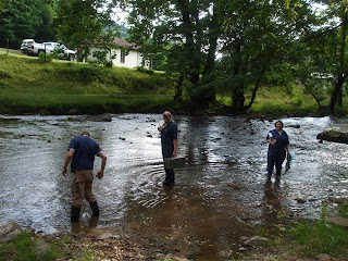
I apologize for the delay in posting, but I've been absorbed in reviewing everything I've learned over the past 3 years on cattle, horses, pigs, sheep etc because I am on my ambulatory large animal medicine block. That's right folks, I'm doin' the James Harriot thing for three weeks. I've already completed the first week and have been delightfully surprised. I initially came in with much anxiety and concern about the danger of working with the large animals and the smell that comes with them. However, my first week involved working with exclusively cattle (both beef and dairy) and I had a blast. The clinicians are fantastic, the clients are real characters and most importantly, you're outside and driving around everywhere! Speaking of which, the picture shown here is of my classmates and I returning to the truck after cutting the teat off of a lactating beef cow that had severe mastitis in one of the quarters of its udder. Note, there was a bridge we could use, but since we were wearing coveralls and big rubber boots, what fun would there be in doing that. :)

I do admit that I am very fortunate to be attending school in such a beautiful area of the country. While driving around, the views of the Appalachian mountains still never cease to amaze me. Initially, however, I was a bit concerned because some of the calls have been to farms out in....how shall I say......Deliverance country. Every client that I've met has been great to work with and very friendly. I'm trying to diet at the moment, and these farmers are making difficult by offering us their delicious home-made country foods, and it's considered an insult to turn it down. As mentioned previously, the food animal clinicians are really nice, very willing to teach, and very down to Earth. There's one clinician in particular that I've worked with alot this past week, and I'm grateful to him for his patience with us small animal folks, and for the memories that will last with me forever. In addition, he goes to Hardee's every single day for lunch (no joke) and somehow remains in fantastic shape.

On my first day of the block (first call actually), we were called out to this beef cattle farm about an hour south of the school to help pull a calf out of a cow who should've been out two days prior. Before I go further, I need to point out why I said "beef cattle". Dairy cattle spend their whole lives being handled and spending alot of time indoors, so they are much, much easier to work with; beef cattle however live out on pasture their whole lives and you have to herd them into a chute to work with them....and they aren't too fond of the idea either. The farmer claimed that the cow trusts him and won't run away if its just him, so we gave the farmer a syringe with sedative in it to give the cow (which he did with no problem), and we went to go check on it in a few minutes. We offroad through the pasture to where the cow was laying on its belly, thinking to ourselves that the sedative worked. The clinician hopped out of the truck and got a rope ready to lasso her head and try to tie her to a fencepost long enough to put a halter on her. Before doing so, the clinician told me to hop in the driver's seat of the truck and get ready to drive it, and I had no idea why. I quickly learned why as the cow lept up after getting lassoed and started to take off with the clinician holding on and running for dear life shouting at me, "Drive the truck to me so I can tie her to it!" While I do know how to drive stick, I've never driven a big pick-up truck before, nor have I driven off road on Appalachian mountain foothills at a 60 degree angle. The clinician is actually a really big guy, easily 6'5" and built like a house, but the cow couldn't have cared less. So here I was driving this truck around the pasture after this cow, and I guess fate pitied us because the cow ran indoors where the clinician could tie her to a wall.

Thankfully, we were able to save the cow because we could take the calf out without much difficulty or surgery, but the calf was long dead. This was the first time the cow had given birth, and sometimes they just produce calves that are too large for them to come out naturally. I still have plenty more stories, but I'm going to stop here for tonight because I can go on for while, but I will be doing my best to post each day this week to catch up on all my stories. Thanks for reading!

Friday, June 15, 2007

A Gallery of Craziness

Well, my external block is over and I had one helluva time. I will post a detailed description this weekend, however, I promised to post some pictures I took of my cases. Also, I failed to post a reply to comments on my previous posts because of my busy week, but I wanted to say thank you Elaine for reading my blog and for your comments. It reassures me to know there are others who feel the same way as I about birds :). Now, onto the pics! (Don't worry, I am refraining from showing the gross/graphic ones I took from either necropsies or surgeries)

This is an English Bulldog with a disease called "Seasonal Flank Alopecia": a disease where dogs will lose hair from their sides for reasons unknown (not allergies), and it does not itch. The hair usually grows back.


These are the remains of a corn cob that were taken out of a dog's stomach via exploratory gastrotomy. The owner's were not neglectful or anything, the dog rumaged through their trash while they were away. I (jokingly) wanted to say to the owners, "I hope you enjoyed the corn, because that was the most expensive corn cob you'll ever have."


Here is a leopard gecko that suffered some serious burns along its back. My initial thought was that the owner was using a heat lamp that was too strong, however the reptile expert at the hospital thinks it was potentially due to too much UV light.


For those of you who have never seen one of these, this is a Sphinx cat, a hairless breed. While their appearance may not appeal to everyone, they have fantastic personalities and (from what I've heard) good for people who are allergic to cats. However, they have a plethora of health problems, like difficulty developing a proper immune system. This one in particular had pneumonia.


This is a xanthoma (a common benign tumor of birds) on the wing of a parakeet. In this case, the tumor was so large that the bird not only couldn't fly, but it couldn't remain on perches because it would fall off from the weight. The bird went to surgery and the mass was removed, but because of extensive local invasion, it will likely recur.


Here is a young boxer dog that presented with a huge abscess (mentioned in my previous post) under his jaw. The abscess was not removed in its entirety because the post-operation care is rather intensive. We just lanced it open, drained it, and placed a drain in it to keep it as small as possible till the owners returned home to NC where the full procedure should take place. This dog was a really great patient and incredibly sweet.


This here is a tumor with a bird attached to it. We made a valiant effort to take this bird into surgery and have it taken out, but sadly, the bird did not make it through surgery. I will be taking a biopsy of the mass back to the vet school to find out what type of tumor it is.


I apologize for the quality of this picture, but here is a parakeet that was brought in from a Petsmart because its right leg was just a dead husk, and thus completely useless. We have no idea what the inciting incident was to injure the leg, but it had to go, so the leg was amputated.


This one is for Dave, who requested more cute pictures. I will leave it to The Foodist to insert appropriate and hilarious captions :). I hope you enjoyed the pics!

Monday, June 11, 2007

On a More Positive Note....


See? I told you my next post would be mostly a positive one. It's hard to get more positive than a bulldog puppy, or as I like to refer to them, cuteness in its purest physical form. The pup was only visiting the hospital to get its next round of vaccines, but I still demanded to take the time to see/play with it. I mean, come on.....wouldn't you?

Onto things that I've worked with over the past week. A young boxer came in with a huge abscess under and behind it's jaw (it was about the size of a grapefruit). The kicker though for this scenario is that the owners were a young couple from NC just visiting for a relatives high school graduation. Just over the course of this weekend, that abscess grew to the size that we observed. The vet recommended that the walls of the abscess be taken out down in NC, but still offered to open it up and place a drain in it to help prevent it from growing. We did as such and as soon as the dog was awake from anesthesia, we could tell it was feeling a lot better. It was amusing watching the vet teach the clients how to replace a bandage over the dogs neck that involved using a baby diaper.

One of the more amusing appointments I saw involved a morbidly obese Golden Retriever. The owner was a great, older gentlement (my guess was late 70's) who was a real card, and a pleasure to work with. While I was checking in the appointment before the doctor visited, he asked me why she was panting so heavily all the time. I tried to be professional and politically correct by responding with, "Well, we'll see what the doctor thinks, but it may have something to do with her size." Later, partway through the doctor's exam of the dog, the client said, "Yeah, I asked your student here why she's breathin' so heavy and he said it's cuz she's FAT!" Thankfully, the doctor just assumed I didn't say it like that because I wasn't about to correct the client, especially since he was saying it in a jovial manner.

This past week I was able to work with a number of different birds: a couple parakeets, a couple African grey parrots, some cockatiels, and a conure. I love working with the African greys because they like to talk to you while you handle them (even though they aren't readily handled). While I found it fascinating learning how to work with birds, my experiences at this clinic just further reinforce how I will most likely avoid them entirely post graduation. For those of you who have never owned a bird, they are by far and away the most frail creatures on this planet. Not only can you easily break their bones while removing the metal rings around their legs (or just from handling), they can literally die just from the stress of handling. The latter happened last week. A bird was brought in because the owner noticed it got its leg caught in something in its cage and just wanted to drop it off at the hospital for x-rays and to make sure everything was ok. The bird was left overnight, and the next morning a technician took the bird out to measure its weight. I observed the technician and he was not overly forceful with the bird by any means, but after taking the weight and placing it back in it's cage, it literally just tipped over and died. While the vets do explain to every bird owner how this can happen, the phone call still has to be made to the client. To be quite honest, it's not one I'd like to make.

Thanks for reading, and stay tuned as I post a bunch of pictures from my time at this clinic!

Sunday, June 10, 2007

A Wonderful time

Well, I'm two-thirds of the way through my second block and I'm absolutely loving it. This hospital has me working between 10 and 12 hours a day, and for once....I don't mind. In fact, I hardly notice the time go by at all. Thankfully, I think the owner of the place is really hyperactive which leads to the place being especially busy all the time. The one thing I hate the most at a job, above unsatisfactory coworkers and employers, is being bored. For the time being, the only part of my day where I'm bored is while I'm eating lunch, which is only for 20 minutes anyway.

Probably the highlight of my past week was getting to travel with the other doctors at the practice to this local meeting of a group called the DC Academy of Veterinary Medicine. The first Thursday of every month, doctors from all around DC gather for this monthly conference where speakers from all over will present the latest developments in the profession. This month, the speaker was a professor from NC State who presented the latest information about tick-borne diseases in dogs and cats, such as: Babesiosis, Ehrlichiosis, Lyme's Disease, and Rocky Mountain Spotted Fever. Not only was the speaker great, I especially enjoyed his talks because I knew I wasn't going to be tested on the information ;). There must've been a few hundred veterinarians there who all really reinforce what a great community this profession provides. Oh, and best of all.....free breakfast and catered lunch ;P.

On a sadder note, I assisted with the first few euthanasia's at the hospital since I've been there. One was done on an old Golden Retriever who had malignant melanoma and prostate cancer to boot. The vet asked me to join him in the room and warned me about what a tear-jerker is was going to be. In the room was not only the dog, but the entire family consisting of a husband, wife, and two teenage daughters, both of which were audibly bawling. By the end of it, the vet started losing it as well since these clients have been coming to him for over two decades. I managed to keep my composure, but only because of my own awkwardness. To be completely honest, I never have any idea of what to say in these situations so everytime I've assisted with a euthanasia, I've kept my mouth shut. While I did my best to take the doctor's consoling words away with me, it wasn't exactly the type of scenario where it would be appropriate to take notes. The other euthanasia I had difficulty with involved an owner who couldn't bear to watch the act, so they left the dog at the hospital. I don't personally understand how someone could view it that way, I still certainly respect it. This dog was a lab who had a really distended abdomen, was having some difficulty getting up, and was defecating all over the owner's house. What bothered me though was how the dog still seemed happy and had a positive attitude. Fortunately, the owner opted for a necropsy for some closure, which I performed and found the dog had hemangiosarcoma of the spleen: a really nasty malignant tumor which can (and did in this dog) spread to the heart, and can burst at any point causing the dog to bleed to death internally. Therefore, while this dog did seem happy, he could've dropped dead in a terrible way any day.

This post is getting a little winded, so I'll post other cool cases (mostly positive ones) I saw last week in a post tomorrow. Thanks for reading!

Sunday, June 3, 2007

Onto the Living

Well, one block down, 16 to go. I completed my lab services/necropsy block two fridays ago, and have spent this past week doing an externship at a hospital in Maryland. As the title of this post points out, this is my first block working with live animals, and it is much more refreshing (in regards to both enjoyment and smell). The hospital that I'm working for is absolutely astonishing. Every other one I've either visited or worked at had a number of personnel that were absolutely miserable, whether they be receptionists, technicians, or even the vets. While I haven't met all of the employees of this establishment yet, every single person I have met has a great time and has a wonderful outlook on their job. You won't find many places where every single employee is actually having fun. In addition, all of the doctors and technicians are understanding of how this is only my second block and the fact that I have minimal clinical experience prior to entering vet school. All of them are superbly patient with me and love mentoring me.

In just one week, I have learned a plethora of new information that it's fruitless for vet schools to attempt to teach. The bonus in this case is how this clinic sees alot of exotic animals, which I have gained an interest in during the course of school. Thus far this week, I have seen and worked with an iguana, a burmese python, a bearded dragon, a cockatiel, 4 guinea pigs, 2 ferrets, a hamster and a rabbit. There are so many little things about each species that can really only be taught through a mentor, such as handling techniques, how to perform physicals, etc. Thankfully, I purchased a PDA during my last block so I've been taking down as many bits of wisdom that I can. Here are some examples:

-When force-feeding reptiles, make sure you look into their mouth to ensure that they don't aspirate any food into their lungs since reptiles don't have a cough reflex. If they do aspirate, they'll simply just die in a day.

-When handling a rabbit, always support their rear end, otherwise they will flail their hind limbs and can potentially break their back in the process. That I actually knew, but I didn't know that you should always put them back into their carrier backwards for the same reason. If you put them in facing the inside of the carrier, they'll push too hard trying to get inside.

-I knew that ferrets are more prone than other species to get severe reactions to vaccines, so never ever give them more than vaccine at once.

-If a female guinea pig presents with hair loss without being itchy, it is most likely due to an ovarian cyst.

-If a dog/cat has had immune-mediated hemolytic anemia/thrombocytopenia (where your own immune system starts to destroy your own blood cells or platelets) in the past but recovered, you shouldn't vaccinate them anymore for fear of re-stimulating the immune system to go nuts again. This will require writing a letter to the "man" saying this animal can't be rabies vaccinated.

-"Red-eye" is a common initial sign for glaucoma

There were plenty of other things for me to experience and observe. I watched a dog undergo exploratory surgery and have its stomach opened because it raided the owner's trash can and ate an entire corn cob. Later in the week, I watched an orthopedic procedure to fix a dog's knee (to be specific, the dog had patellar luxation). Above all, I'm really enjoying enhancing my technique with taking and interpreting radiographs (aka X-rays). While we are taught radiology very well in lecture, the best way to learn how to read a radiograph is through practice. So all in all, this hospital is giving me more practice and knowledge than I would've ever imagined, and in just one week. I hope to post more stuff as I see it, so long as there is time to post :).

Tuesday, May 22, 2007

Mixed Feelings

For those of you who know me personally, you are well aware of my concern for the future of animal legistlature. Many people are in the dark about how much animal law is growing in this country; in fact, there are a number of law schools that offer an entire course on animal law. My greatest concern is with that of the Guardianship Campaign. There is a well-supported push across this country to legally promote the status of companion animals from property to wards. While this may seem insignificant, this would mean that when suing someone over the life, health, or welfare of their companion animal(s), they can receive non-economic damages (i.e. emotional damages) from their suit. Personally, I am really torn about the whole issue. For the most part, I can understand the purpose of the campaign and agree how we in the United States have certainly elevated the status of pets from their utilitarian purpose to that of family member. Being a veterinarian in training, I should appreciate this more than most. On the other hand, this will result in some serious economic ramifications that many don't realize. My theory is that once this becomes nationwide, there will be an excessive number of these lawsuits and the law will be greatly abused. Just go here to understand where I'm coming from. Most importantly, I feel there will be a great change in the costs of small animal veterinary services. With so many people becoming sue-crazy, there's not a doubt in my mind that malpractice insurance costs for veterinarians will skyrocket to match those of human physicians, thereby causing veterinary services to likewise skyrocket in cost.

Again, I will point out that this movement is for a good purpose and it has already been used to great effect. A professor I had in my first year told me about a case where a woman in Kentucky sued a ranch over her two appaloosa horses. The owner was a professional and traveled for months at a time for her job, while boarding her horses at this ranch. She came back after a month of traveling to find that the owners of the ranch had to send her horses to some other ranch they owned on the far side of the state. Concerned, she drove all the way to the address given to her, only to find that the place didn't exist. After hiring a private investigator, she found out that the ranch owners sold the woman's horses to one of the last remaining horse slaughtering facilities down in Texas, to be sold to the French, Canadians, etc. She, of course, sued the ranch owners and received an enormous cash settlement. I also see the the guardianship changes having positive results in reducing the number of malicious poisonings of pets across the country. I don't know the statistics, but veterinarians everywhere see dogs and cats that have been poisoned with ethylene glycol (antifreeze), rat poisons, insecticides etc. At the moment, when you sue someone for mistreatment of your animal, all you can be awarded is the property value of the pet. So to put it in perspective, sueing over an 18 year old diabetic cat isn't worth the hassle. What are your thoughts on the whole campaign? I'm interested to see what other people think and see if they have the same concerns.

(In regards to my work, I'll post again probably this weekend. There is an exam for the whole lab services block this Friday that I have been studying for all this week, preventing me from posting in much detail. Stay tuned!)

Tuesday, May 15, 2007

One week down, 51 to go

Well, so far so good. My first week (last week) went very well. So far I've done a necropsy on a foal (the one described previously), an adult horse with Ehrlichia and possibly Equine Protozoal Meningoencephalitis, a pig, and a goat (I don't remember what the latter two had wrong with them). I'm beginning to get used to the smell, but there are still some that are giving me problems. Without question the most interesting case thus far was another horse that needed a necropsy yesterday. Because of some work being done to the roof of the large animal facilities at the vet school, all of the patients had to be moved over to a research barn for a day or two. Go figure a patient is placed over there who later became suspect of having a disease caused Strangles: a highly contagious disease where a bacteria (Streptococcus equi) causes many problems inside the horses body, but most notably invades the lymph nodes right behind their jaw on each side, resulting in severe enlargement of those nodes. This often results in difficult breathing and eating, amongst other systemic signs. The students and clinicians isolated the horse and treated it appropriately for Strangles, and it appeared to begin improvement, but at 4:30 AM yesterday the technicians found it dead in it's stall with blood everywhere.

Because of the bizarre nature of this case, the horse was transferred to us on necropsy. We let the pathologist on duty do the honor of dissecting the head because it requires experience and precision when dealing with this disease. Upon opening the head and examining the throat area, the pathologist found a 5 cm long piece of metal wire that the horse had ingested that forced it's way back up into it's head, resulting in an infection with an abscess that resulted in the enlarged lymph nodes. To explain the bleedout, one must be aware of a lil anatomy. Horses (for whatever reason), have these two empty spaces inside their head just below their brain, but behind their throats called Guttural Pouches. These can be infected with many different infectious diseases as a separate entity, but the wire made it's way inside the right pouch. More importantly, right behind each pouch is the internal carotid artery, the primary blood supply to the brain. The carotids are under extreme pressure, so when lacerated by say...oh....a wire.....the horse (or any mammal for that matter) can die of blood loss and lack of oxygenation to the brain in a very short period of time. Overall cases like this are very rare in horses because they are selective, careful eaters. Usually cases of eating foreign bodies are reserved for cattle....or as I call them, the dogs of the large animal world :).

On a completely different note and per request from a previous post, I'd like to tell the story about how I got my cat, Kahlua. Last fall, when my classmates and I were first learning surgery, there were a total of three surgery labs over the course of the semester prior to that practical final I mentioned before. Two of these labs were spays on dogs, and one on cats (the surgeon that day gets to do both a spay and a neuter). The staff just assigned who was doing which surgery and they asssigned me to the cat procedures. All students are required to go to the school at 5:00 PM the night before the surgery to do a pre-surgery physical exam, do bloodwork and urinalysis to ensure the cats/dogs are satisfactory patients for anesthesia. Upon examining my patients, I found that the male was perfectly ok, but the female was another story. She was depressed with a heart rate of 240 beats per minute (200 is the most you can accept, and even then it's only if the cat is scared or excited), had difficulty breathing because of a mucous discharge coming out of her nose and eyes, had a heart murmur, and a temperature of 103.5 F (101-102 is normal). My partners and I kinda looked at each other with confusion and hesistation, so we decided to call over the surgeon on duty to take a look at the cat. His response was "Oh this cat's fine, just check the temperature in the morning just to be safe." Doing as we were told, we repeated the entire exam the next morning to find that nothing had improved, and the tempurature was up to 105.1 F. There was no surgeon to be found anywhere, so we just explained our concerns to a technician who said she'd leave a note for a surgeon once one came in for the day. After three hours of lectures in the morning, I came down to find out that the sickly cat had been taken away and replaced by a beautiful Birman cat named "Mese" because the rescue group thought she looked like a siamese but with long hair (and to be honest with you, I would've made the same mistake b/c I've never seen a Birman in person before). I was then forced to skip my 11:00 lecture to hurriedly fill out all the necessary paperwork and perform all the stuff done to the previous cat the night before. I say hurriedly because I still needed to eat lunch and all animals are expected to be anesthetized by 12:30. Thankfully, "Mese" was friendly and in perfect health. My groupmates joined up with me shortly afterwards and I performed my first surgery without complication.

Looking back, there's not a doubt in my mind that the previous cat would've died on the table without me even cutting it. From what I heard, it recovered and was spayed successfully at a later date. All of the dogs used in the surgery are owned by local clients, however the cats are all from a rescue organization and need homes. I saw that Mese needed a home, but found out that there were 14 other people interested as well. Being a vet student and the one who spayed her, I was bumped up on the list as far as possible, but they had already started the interviewing process for the first prospective adopter. I waited a couple days, then was contacted by a fellow student who said the interview didn't work out with the person because neither she, nor any of her references could be contacted after repeated attempts. So after much anxiety, I was selected to get her as my own, my very first every surgery patient. In fact, as I write this she is laying across my lap, making typing very difficult. She's a friendly, beautiful cat who thankfully doesn't hold it against me how I took away her womanhood ;). I don't have a good picture of her at the moment, but I'll take one and post it here this week.

Monday, May 7, 2007

The die is cast...

Well, it's a done deal. I am now officially a senior student gettin' my learn on :P. The first day was both exciting, strange, and depressing all at once. Depressing because the now graduating seniors had to come by the school today to transfer over their cases to us before 8 AM, then proceeded to drink in the parking lot, while cheering/taunting myself and other classmates as we approached the school. Never once have I felt such a strong sense of jealously in my entire life. Strange because it's odd to now see myself and my classmates wearing the navy blue clinician's jackets and running around like crazy people instead of it being a mere aspiration. Exciting, of course, because this is why I came to school and how 90% of what is learned/retained happens this year.

My first block is in "lab services" which, thankfully, is a good transition block into 4th year because it is one of the more laid-back services in the school. Because the pathologists, bacteriologists, parasitologists, etc never directly deal with clients, we don't have to abide by a true professional dress code (which is nice for me since I despise wearing ties :P). Each day consists of reviewing concepts and techniques from classes such as bacteriology, toxicology, virology, parasitology, clinical pathology etc all morning; then the afternoons consist of performing necropsies on deceased animals. Today, we spent the entire morning looking at fine-needle aspirate cytologies (where you just stick a needle into a lump, shoot the contents onto a slide and look at it under a microscope) and reviewing how to differentiate normal tissue from an inflammatory process or even cancer. We even talked about how to identify certain types of cancer with this diagnostic tool. Personally, I am very thankful for this since almost all classes thus far have talked about how to read and diagnose biopsies, which your general practicioner never does and just sends off to a lab. Fine needle aspirates however are easy, cheap and readily done in private practice.

This afternoon however was when things got really interesting as I performed my very first necropsy. It was done on a 9-day old Thoroughbred foal who, most likely, died of failure of passive transfer. In all mammals, a neonate acquires some immunity from its mother to keep it protected for the first few weeks of life until its own immune system develops and begins to function. In humans, dogs and cats this is done while still a fetus inside the mother. In cattle, horses, sheep, goats etc however, this occurs through ingestion of antibodies in the milk. Unfortunately, these antibodies in the milk are only present for the first few days of the animal's life so, if for any reason the animal does not nurse, it is left without an immune system for a few weeks. This foal in particular had a severe infection that made it's way into the bloodstream (which is real bad news), and resulted in some severe lesions around its lungs, in its stomach, and in its intestines. I had to think to myself, "Now this is how you start a senior year" as I was hacking away inside the chest cavity. I say this in jest because this was a very disgusting process. There were a few gallons of fluid inside its chest that I just had to slosh around in while removing the lungs and heart (both of which needed to be removed for microscopic examination). Not all animals have such distinct lesions so while of couse it was bad for the horse, it was good for us since it helped us reach a diagnosis.

I apologize for the gross details; they won't really continue after this block is over....hell, I'll probably get too grossed out talking about them. Needless to say, I'll probably become a vegetarian until this block is over. :)

Wednesday, April 25, 2007

So Close Yet So Far

I want to apologize to all of you who read my new blog. It would appear to me that I created it a little prematurely. While I do state in the "About Me" section that I am a 4th year veterinary student, I won't officially be until May 6th. I've been uber-busy cranking out my last assignments and finals of the semester/my life, then I will be away for the next week on my miniature summer vacation. Afterwards though I shall be rocking the vet medicine 1st hand and will be posting all my triumphs and hardships quite frequently. Stay tuned! :)

In the meantime, I try to keep up to date on what matters in the animal world and will post stuff on here every now and then that I find interesting. Take this article for example which highlights a critical issue in today's animal world :P. To be honest, I just assumed cockfighting was illegal everywhere; that is, until I came to veterinary school and had this question on an exam in my first semester.

"Cockfighting is legal in Virginia so long as there is no wagering or gambling of any form associated with the event." True/False

At that point in time it was legal in Virginia, and I can proudly say that I will be one of the last people who can say they had an exam question about the legality of cockfighting. My favorite part of the article above is the veterinarian talking about the argument against banning the "sport". Part of the culture? Get out of here. If anyone here from Louisiana or New Mexico happens to read my blog, can you validate this? or on the other hand, further the doctors belief that "cultures" change?